This article is for educational purposes only. It is not financial advice, not legal advice, and not tax advice. Figures vary by individual circumstances, so consult a qualified professional before acting.
Here's the mistake I keep watching smart clinicians make: the AI spits out a number, the number has two decimal places, and it goes straight into the report. Done. Trusted. Filed.
Don't do that.
I recently watched a follow-up chest CT nearly trigger an oncology workup because an AI tool flagged a pulmonary nodule at 11.2 mm, up from 8.4 mm six months earlier. Alarming growth, right? Except it wasn't growth. The algorithm had grabbed a different slice and folded part of an adjacent vessel into its outline. Ten minutes of manual review showed a nodule that hadn't changed at all. The "progression" was a segmentation artifact, and the number was already sitting in the draft report.
That's the core idea of this article: AI radiology measurements can be genuinely useful, but the first number is often an early estimate. A first pass. Not a validated final measurement.
When I talk about measurement integrity, I mean four things working together: image quality, segmentation reliability, unit and scale correctness, and correct anatomy/ROI selection. If any one of those fails, the number fails, quietly. Ahead, I'll walk you through the failure modes I see most (wrong slice, ROI drift, scale misreads, post-processing artifacts) and the red flags that should stop your hand before it types.
What the "First Number" Usually Represents (and Why That Matters)
An AI measurement doesn't appear by magic. It travels a pipeline, and errors can enter at every single stage: the DICOM series comes in, the data gets normalized, the model proposes a region of interest, segmentation runs, and then a measurement gets computed from pixel geometry. Only after that should quality checks kick in, boundary confidence, slice-to-slice consistency, spacing verification. The problem? What pops up on your screen may be the output from before those checks finished. Or before they ran at all.
Three different values can exist for the same finding:
- Raw prediction, the model's first guess, straight off the segmentation.
- Post-processed measurement, smoothed, snapped to rules, QC-filtered.
- Clinician-adjusted final value, the only one that belongs in a report.
Guess which one many interfaces show you by default. The earliest. Because it's fast, and speed sells software.
The mistake to avoid: assuming the first number is already calibrated to correct pixel spacing, orientation, and slice selection. It frequently isn't.
Red flags that scream "preliminary output":
- Suspiciously precise decimals (an 11.27 mm nodule on a 2.5 mm slice? Please.)
- Abrupt jumps in size between adjacent slices
- Implausible anatomy-to-size ratios, a "3 cm adrenal nodule" that's bigger than the gland
- Overlays that visibly ignore the lesion's actual boundary
Myths That Get Clinicians Into Trouble
Myth 1: "The AI value is already final." Reality: it may be a first pass or a UI default that hasn't cleared QC. I've seen workstations display the raw number while the validated one sat one click away. One click nobody made.
Myth 2: "If it looks precise, it must be accurate." Reality: precision is not correctness. A segmentation error can produce a beautifully confident, totally wrong measurement. I call it precision theater, decimals performing reliability they haven't earned.
Myth 3: "AI is objective, so image quality doesn't bias it." Reality: garbage in, garbage measured. Low contrast, motion, streak artifact, protocol mismatch, all of it skews segmentation. The model doesn't know the scan is bad. You do.
Myth 4: "It agreed with me once, so it's reliable." Reality: performance drops with rare morphologies, unfamiliar scanners, atypical anatomy, post-surgical changes. Yesterday's validation means nothing for today's weird case.
Here's the Mistake Avoider rule, and I'd tattoo it on the workstation if I could: every AI number earns trust through verification. Not vibes.

Reality Check: Common Failure Modes Behind Wrong Measurements
Failure mode 1: Wrong slice, wrong level. The AI measures its most confident slice, not your clinically intended one. You wanted the longest axial diameter for RECIST; it measured the slice where the lesion looks biggest and cleanest. Those are not the same thing.
Failure mode 2: ROI drift. The segmentation slightly over- or under-shoots the boundary. For a diameter, that's a modest error. For a volume, it's brutal, the math amplifies it. A measly 5% error in radius on a spherical lesion barely moves the diameter but swings the volume by roughly 14-16%. Small boundary, big bias.
Failure mode 3: Scale and unit errors. Pixel spacing problems, mangled DICOM metadata, magnification factors on plain films, mm reported as cm. Rare, but when it happens, it's catastrophic and completely invisible unless you check.
Failure mode 4: Confounding anatomy and artifacts. Surgical clips, embolization coils, calcifications, motion blur, partial volume effects. I've watched an algorithm confidently outline streak artifact from a hip prosthesis as if it were tumor. Confident. Wrong. Dangerous.
Failure mode 5: Protocol mismatch. A model trained on thin-slice arterial-phase CT will not behave the same on your 5 mm portal venous reconstruction. It doesn't warn you. It just quietly underperforms.
Actionable red flags, treat any of these as a stop sign:
- Sudden measurement jumps between serial studies
- Numbers that defy expected growth patterns (a nodule "doubling" in three weeks)
- Segmentation boundaries that visibly leak into adjacent tissue
- Low or missing confidence indicators
- An output that answers a different question than the one you asked
How to Verify the AI Measurement Without Becoming Slowed Down
Now the opposite mistake: turning verification into a five-minute ritual that collapses under real workload pressure. That's how you end up with checkbox medicine, going through motions, catching nothing. The goal is sixty seconds of high-yield checks. Here's the sequence:
Step 1, Confirm the question. Longest diameter? Volume? Area? Make sure the AI is computing the metric the clinical scenario actually demands. A beautiful volume is worthless if the tumor board needs a RECIST diameter.
Step 2, Check the geometry. Verify slice level and plane orientation. Does the overlay sit on the structure you intended, on the slice you intended? Scroll one slice up and one down. Takes four seconds. Catches the wrong-vessel problem every time.
Step 3, Validate the scale. Glance at the units and pixel spacing shown in the viewer. If the metadata looks off or the conversion is unclear, distrust everything downstream. Millimeters are not a detail.
Step 4, Assess segmentation quality. Does the boundary hug the lesion? Is it consistent across slices? Does the ROI leak into adjacent tissue or exclude the spiculated edge that matters?
Step 5, Apply trend logic. Pull the priors. AI is not exempt from biologic plausibility. If the number implies growth that makes no physiologic sense, the number, not the biology, is probably wrong.
Step 6, Respect the QC signals. If the model itself reports low confidence, believe it. Treat the value as provisional and measure manually.
Notice what this flow does: it routes bad numbers to a human fast, instead of letting them coast into the chart on momentum.
Reporting Safely: Wording, Uncertainty, and Documentation Pitfalls
Don't write AI numbers as facts. Write them as what they are: measurements with a method. Document whether the value was AI-derived and whether you verified the overlay. "AI-assisted measurement, segmentation reviewed" costs you six words and buys real protection, clinical and medicolegal.
When image quality is borderline, say so: "Measurement may be limited by partial volume effect and motion artifact." That sentence isn't weakness. It's honesty that keeps the next clinician from anchoring on a shaky number.
And beware your own brain. Anchoring bias is real: once you've seen 11.2 mm, your manual measurement mysteriously drifts toward 11. Verify before you look at the value when you can. At minimum, be willing to override it.
Standardize the workflow. Capture the model version and QC status when available, and always note when manual correction was performed. The documentation error that makes me wince hardest? Reporting an AI output with the wrong metric, volume quoted where a diameter was requested, or no units at all. That's not a typo. That's a patient safety event in waiting.
When to Override or Escalate: Hard Stop Scenarios
Some situations aren't "verify quickly" situations. They're hard stops. Never rely on an AI first number when image quality, ROI integrity, or metric alignment is in doubt. Full stop.
Escalate to manual measurement or expert review when:
- The model reports low confidence or failed QC
- The segmentation boundary is visibly wrong, trust your eyes over the overlay
- Severe motion or streak artifact contaminates the field
- Post-surgical hardware (clips, coils, prostheses) sits anywhere near the target
- The anatomy is atypical: congenital variants, distorted post-operative beds, rare morphology
- The measurement contradicts the physiologic pattern of prior studies with no plausible explanation
And think beyond your own reading room. Protect patients at the institutional level: define which confidence scores trigger mandatory human review, set thresholds for when a model needs recalibration, and retrain when scanner fleets or protocols change. A model validated on last year's fleet is a hypothesis about this year's.
Bottom Line: Treat AI Measurements as a Second Opinion, Not a First Answer
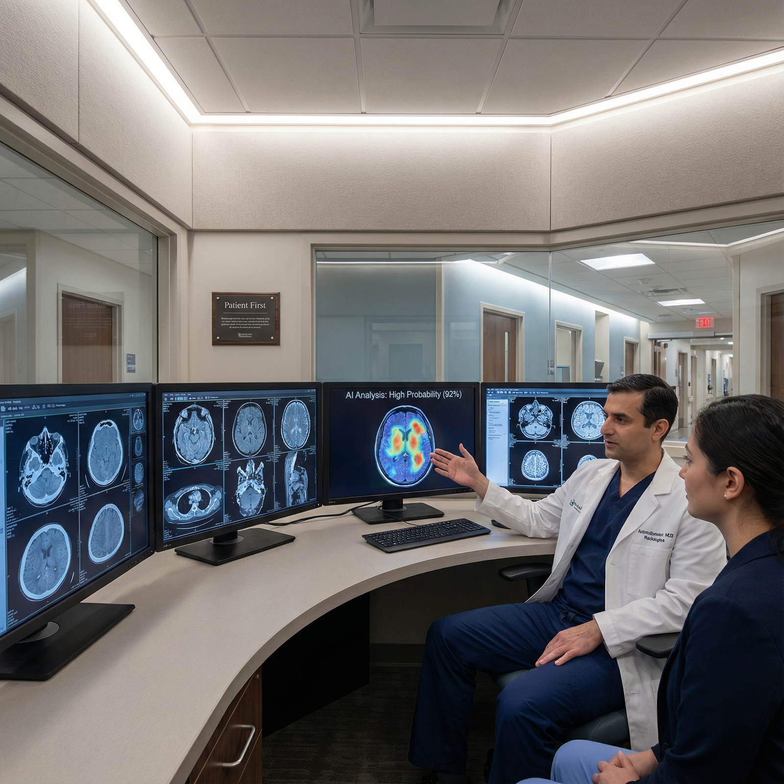
The protective message, one more time: don't trust the first number. Verify it against the clinical question, the image, the scale, and plain biologic plausibility. AI is a fast, tireless second reader, a genuinely valuable one, but a second opinion you check, not a first answer you obey.
Pin these to the workstation:
- Never treat the first displayed measurement as final. Verify metric, overlay, slice level, and scale before reporting.
- Most AI measurement errors are boring, not exotic. Wrong ROI, wrong slice, wrong units. Confidence signals and prior-trend plausibility catch them early, if you look.
So here's your call to action, and I'm serious about the "today" part: commit to one process change before your next shift. Make it this one, before any AI-derived number enters your report, you have personally confirmed the slice level and the units. Two checks. Ten seconds. That's it.
The first number is free. The verified number is what your patient deserves.