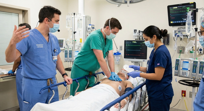
It's 2:30 a.m. Your pager vibrates off the nightstand. The ED nurse is on the line: a patient's IV infiltrated during a CT angiogram, and now 80 mL of contrast is sitting in their forearm. The patient is anxious, the arm is swelling, and the nurse wants to know what to do next.
Welcome to overnight call.
Contrast extravasation isn't just a blown vein. Hyperosmolar contrast agents are toxic to subcutaneous tissue. If you mishandle this, you're looking at skin necrosis, compartment syndrome, or a lawsuit. Your goal right now is simple: rapid assessment, stop the exposure, protect the tissue, escalate appropriately, and document everything.
"Good on call" doesn't mean you have all the answers memorized. It means you stay calm, execute immediate actions in minutes, and make follow-up decisions within the hour. Here is exactly how to handle this call, step by step.
This article is for educational purposes only. It is not financial advice, not legal advice, and not tax advice. Figures vary by individual circumstances, so consult a qualified professional before acting.
Step 1, Confirm What's Happening (Extravasation vs Other IV Complications)
First, make sure you're actually dealing with extravasation and not a simple hematoma, phlebitis, or an allergic reaction.
Listen to the nurse's report. The classic clues are burning or pain at the IV site, visible swelling, coolness or pallor of the skin, and a slowed or absent infusion. If the pump is alarming "occlusion" but the bag is empty, that's a massive red flag.
You need to differentiate this from other complications early. A simple hematoma is localized and firm. An allergic reaction presents with systemic symptoms like wheezing, hives, or hypotension. Compartment syndrome presents with pain out of proportion to the injury and pain on passive stretch. Early distinction drives your urgency.
Get to the bedside. Stop the injection. Stop the pump. Inspect the site. Palpate the swelling. Measure it if you can.
Here is a quick decision-support framework to help you triage the symptoms immediately:
| Finding | Suggests Extravasation? | Action Now |
|---|---|---|
| Pain / Burning at site | Yes, highly likely | Stop infusion immediately. Assess neurovascular status. |
| Visible swelling / Edema | Yes, highly likely | Mark borders with a pen. Elevate limb. |
| Blanching / Coolness | Yes, suggests ischemia | Escalate immediately. Check cap refill and pulses. |
| Blood return still present | Maybe (catheter tip might still be in vein) | Do not flush. Aspirate gently. Follow protocol. |
| Wheezing / Hives / Itching | No (Consider allergic reaction) | Stop infusion. Assess airway/breathing. Treat anaphylaxis per protocol. |
Step 2, Stop the Exposure and Preserve the Line/Access
This is where residents make their first big mistake. Your instinct might be to flush the line to "clear it out." Don't. You will just push more toxic contrast into the subcutaneous tissue.
Stop the contrast immediately. Disconnect the tubing.
If the catheter is still in place, follow your institutional protocol. Most protocols dictate that you gently aspirate residual contrast from the IV hub using a small syringe. Do not forcefully flush. Depending on your hospital's rules, you might need to leave the catheter in place to administer a local antidote, or you might be instructed to remove it immediately. Do not delay treatment while you argue about "perfect" line management. If the protocol says pull it, pull it.
Elevate the limb above the level of the heart to promote venous and lymphatic drainage.
Most importantly, switch to clear communication. Tell the patient exactly what you're doing and why. "The IV fluid leaked into your arm. I'm stopping the infusion, marking the swelling, and elevating your arm to help it drain." This reduces panic and buys you their cooperation.
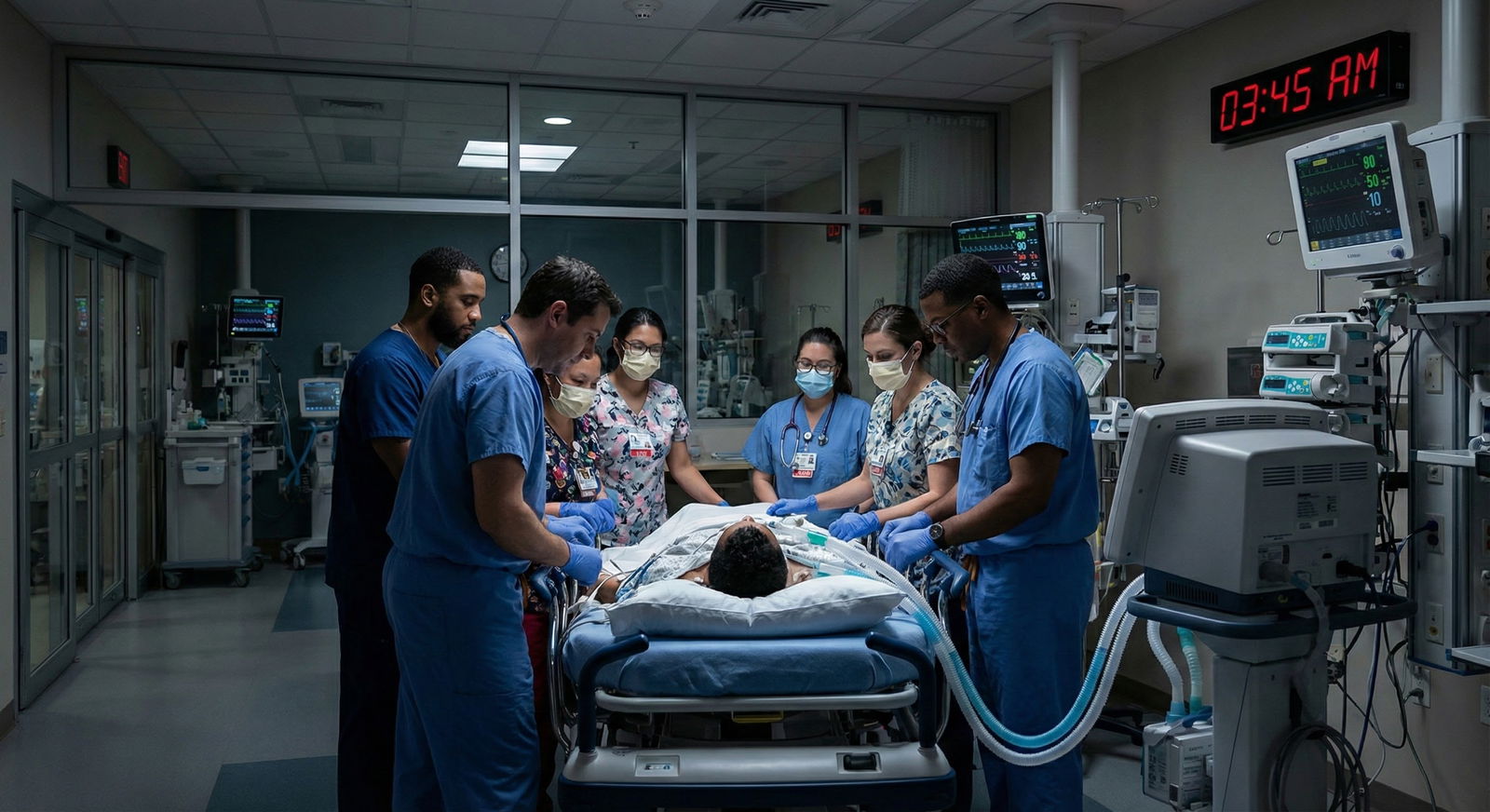
Step 3, Rapid Risk Stratification: Amount, Site, and Agent
Not all extravasations are created equal. You need to rapidly stratify the risk to know if this is a "monitor and wait" situation or a "wake up the plastic surgeon" situation.
Ask the tech or nurse three things:
- How much contrast likely extravasated? (Total volume ordered minus what's left in the bag/syringe).
- What was the agent and concentration? (Iso-osmolar vs. high-osmolar).
- Where is the IV located?
Location matters immensely. High-risk locations include the hands, fingers, near joints, and the dorsum of the foot. These areas have very little subcutaneous fat. Contrast pools right against tendons, nerves, and joint capsules, leading to a much higher risk of functional impairment and necrosis. An extravasation in the meaty part of the forearm is bad; an extravasation in the web space of the hand is a potential disaster.
Assess the severity of the current injury. What is the pain level? Is the swelling rapidly progressing? Is there blistering? Is perfusion impaired? Is range of motion decreased?
Here is your practical escalation rule: If the patient has severe pain, swelling that is progressing quickly, any neurovascular changes (numbness, tingling, weak pulses), or if it's a large volume in a high-risk location (like the hand), you need to call your supervising radiologist or attending right now. Do not wait for the morning handoff.
Step 4, Apply the Correct Local Treatment (Antidote/Adjuncts per Protocol)
Do not improvise. I've seen residents try to massage the site or apply random ice packs because they read a tip on a forum. Stop. Follow your facility's specific contrast extravasation protocol.
Core supportive measures are universally safe and should be started immediately:
- Elevate the limb.
- Apply warm or cool compresses strictly based on your protocol and the specific agent type. (Some protocols recommend cold for hyperosmolar agents to limit inflammation, while others recommend warm to increase absorption. Guessing wrong can worsen tissue damage).
- Apply a dry sterile dressing.
- Avoid compression unless explicitly instructed by a surgeon. Wrapping the arm tightly will compromise perfusion and cause compartment syndrome.
If your protocol recommends specific pharmacological interventions, like subcutaneous hyaluronidase to break down hyaluronic acid in the tissue, or topical DMSO, timing matters. These need to be administered promptly after confirming orders.
Avoid the common pitfalls. Delayed escalation is the biggest one. Using conflicting temperatures (alternating ice and heat) is another. Applying a tight ACE wrap to "compress the swelling" is a massive error that I've seen lead to fasciotomies. Protect the perfusion.
Step 5, Consult Early and Escalate to the Right People
You are not managing this alone. Knowing who to call and what to say separates a smooth night from a chaotic one.
Who to call:
- The radiology attending (or the radiologist on call).
- The supervising service or charge nurse.
- Surgical consult (Plastics, Hand, or General Surgery) only if there is severe neurovascular compromise or suspected compartment syndrome.
What to tell them (Structured Handoff): Use a tight SBAR format.
- Situation: "I'm calling about a contrast extravasation in Room 4."
- Background: "The injection was at 0200. The agent was Isovue 370. Estimated extravasated volume is 60 mL. The IV was in the right antecubital fossa."
- Assessment: "Current pain score is 7/10. Swelling is measuring 15 cm in diameter. Capillary refill is 2 seconds, radial pulse is 2+. Sensation and motor function are intact."
- Recommendation/Action: "I have stopped the infusion, elevated the arm, and applied cold compresses per protocol. Do you want to come see the patient, or should I continue serial exams?"
If you are genuinely worried about compartment syndrome, pain out of proportion, pain on passive stretch of the fingers, tense woody swelling, treat it as an emergency. Escalate to surgery immediately. Do not wait for imaging. Compartment syndrome is a clinical diagnosis.
Step 6, Monitor Like You're Doing ICU Checks (Serial Exams)
The injury doesn't stop just because the pump is off. Contrast continues to draw fluid into the tissue via osmosis. Swelling can peak hours after the initial event.
You need to monitor this patient like they are in the ICU.
Monitoring frequency: Check the patient every 15 to 30 minutes initially for severe cases. Once stable, you can space this out based on your protocol (e.g., every 2 hours, then every 4 hours).
What to document: Serial neurovascular status is non-negotiable. Document the pain trend, swelling progression (measure it with a tape measure), capillary refill, distal pulses (use a Doppler if they are faint), sensation, and motor function.
Watch for delayed complications: Blistering, skin discoloration (mottling or dusky purple), reduced perfusion, and increasing pain that seems out of proportion to the visible swelling.
Set a clear "reassessment endpoint" in your mind. If symptoms are stable or improving at the 2-hour mark, you can down-triage the monitoring. If the pain is worsening and the arm is getting tighter, you trigger your next escalation step immediately.
Step 7, Document Perfectly, Plan Disposition, and Communicate with the Patient
Your medical record is your shield. If it isn't documented, it didn't happen.
Documentation essentials: Write down the exact time of the event, the specific agent, the suspected extravasated volume, the IV site, and the catheter size (e.g., 20g in right forearm). Document the patient's symptoms, your exact exam findings (including measurements), the interventions you performed (including doses and timing of any antidotes), and the names of the people you consulted.
Disposition planning: If this happens overnight, you need to leave a crystal-clear plan for the nursing staff and the day team. Write explicit orders for limb elevation, monitoring frequency, and strict return precautions. Define exactly what triggers an immediate re-evaluation (e.g., "Call resident if pain increases, fingers become numb, or swelling expands beyond the marked borders").
Patient communication: Reassure them, but be honest. Set expectations. Tell them, "Your arm is going to look worse and feel more swollen over the next few hours before it gets better. That is normal. We are watching it closely." Explain what the monitoring and treatments are actually doing.
Quality/Safety step: Complete the institutional extravasation incident report. Do this before you sign out. It's a pain at 4 a.m., but risk management needs it, and it protects you.
Action-First Checklist: What to Do Now (Overnight Edition)
When the pager goes off, don't overthink it. Run this mental checklist:
- Stop the pump. Disconnect the tubing. Do not flush the line.
- Assess neurovascular status. Check pulses, cap refill, sensation, and motor function immediately.
- Stratify the risk. Identify the volume, agent, and location. (Hand/high volume = high risk).
- Start protocol-based treatment. Elevate the limb. Aspirate the line if directed. Apply warm/cold compresses strictly per hospital protocol.
- Escalate high-risk cases. Call the radiology attending for large volumes, high-risk locations, or neurovascular changes. Call surgery if you suspect compartment syndrome.
- Order serial monitoring. q15-30 minute neurovascular checks initially. Mark the swelling borders with a pen.
- Document and dispose. Write a detailed note, leave clear orders for the day team, and file the safety report.
Contrast extravasation is a time-sensitive tissue injury. Stop the exposure, protect the limb, and use your resources. You've got this. Now go answer that page.