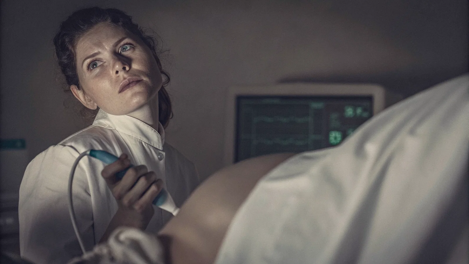
Here's what most residents get wrong: they treat bedside ultrasound like a free answer button. The probe goes down, the screen lights up, and they think they're covered. Then the patient tanks, the attending asks one simple question, "what did you do?", and suddenly the pretty scan doesn't matter.
Program directors know this. They've seen it hundreds of times. The resident who nailed a beautiful lung slide is often the same one who missed a dying patient because they were fiddling with depth settings instead of calling for help. Ultrasound on call can feel like a cheat code, and PDs still coach the same line: know when to move.
What I'll cover here: the wins where ultrasound genuinely shortens time-to-decision, and the moments where PDs wish residents would stop trying to scan their way out of trouble. The goal isn't perfection. It's survival and competence on call when nobody's watching.
The PD Playbook: What "Good Ultrasound Use" Actually Means
Let me tell you what really happens at the program director level. Ultrasound gets graded like communication gets graded. PDs are not looking at your image quality first. They're looking at what you did with the information.
Three grading buckets PDs implicitly score, every time you pick up a probe:
- Question framing. Did you ask a specific, answerable question? "Is there a pericardial effusion?" beats "let's see what's going on."
- Speed and quality of the exam. Can you get the answer in two minutes when the patient is circling the drain? A focused lung exam in under 90 seconds is a learned skill, not a gift.
- Escalation behavior. What did you do when the finding was equivocal, negative, or just plain unexpected? This is where most residents lose points.
The unspoken mindset across every residency program I've worked with is time-to-action. Ultrasound should change what you do within minutes, move the patient, start a treatment, call a consult, activate a protocol. If your scan doesn't lead to one of those within a tight window, you didn't really use ultrasound. You just took a picture.
Here's the uncomfortable truth: a beautiful scan paired with a wrong or delayed plan looks worse than a rough scan paired with immediate escalation when indicated. PDs will forgive ugly images. They will not forgive a resident who sat on a sick patient to optimize their gain.
This is the throughline of everything that follows. Wins live on the side of decision-making. Traps live on the side of image worship.
Ultrasound "Wins": The Situations Where It Saves Minutes (and Sometimes Lives)
A "win" is not "I got a picture." A win is when ultrasound produces actionable results, has a limited training barrier, applies in a high pretest probability setting, and triggers treatments you can initiate immediately. If any of those four legs is missing, you're not in win territory, you're shopping.
Here are the calls where ultrasound consistently pays rent:
FAST in trauma. This is the gold-standard win. Short exam, binary-ish answer (free fluid: yes/no), immediate implications for the trauma team and OR. PDs expect residents to do this in under three minutes. If you can't, you're missing the win.
AAA screening in the right patient. Older patient, abdominal pain, hypotension or syncope? A bedside aorta view is one of the highest-yield things you can do on a night shift. Positive finding = vascular surgery is on the phone in 30 seconds. Negative finding in a low-risk patient is genuinely reassuring. Equivocal finding in a high-risk patient? That's the escalation case, not the reassurance case.
Pneumothorax detection in the right setting. Trauma, post-procedural dyspnea, mechanically ventilated patient with sudden desaturation. Lung sliding present at the apex is a useful rule-out in the correct clinical context. Don't extend that to every dyspneic patient ever, that's scope creep.
Pleural effusion identification. Useful for triage and procedural planning. If you see a big effusion and the patient is short of breath, you've identified a likely contributor and a thoracentesis target. If you see a tiny effusion in a patient whose dyspnea is clearly from something else, you've wasted a probe placement.
Bedside volume status clues (with caveats). IVC assessment is the most over-interpreted ultrasound application in residency education. It's useful as a trend and a piece of the puzzle. It is not a replacement for clinical assessment. PDs know which residents learned this distinction and which ones still quote IVC diameters like gospel.
Cardiac focused assessment (tamponade suspicion). When you see a pericardial effusion in a hypotensive patient, ultrasound just bought you a five-minute head start on a pericardiocentesis or cardiothoracic call. Use it as a trigger for urgent escalation, not a replacement for hemodynamics.
Vascular access and procedural guidance. Real-world upside residents actually notice on call. Fewer needle sticks. Fewer arterial punctures. Faster lines in crashing patients. PDs love this use because it has a clear safety benefit and no scope-creep risk.
The teaching point: "win" doesn't mean definitive. It means ultrasound narrows your differential enough to act, right now, with the team you have, in the room you're standing in.
The Failure Modes: When PDs See Residents Get Tricked
Here's the behind-the-scenes reality most residents never hear: most ultrasound misses are not bad luck. They're workflow errors. Wrong question, wrong patient positioning, poor probe handling, inadequate sweep. The image looked negative, but you weren't actually looking at what you thought you were looking at.
Five failure modes PDs see repeatedly:
The overconfidence effect. You got a beautiful image. You feel covered. So you stop calling for help. This is the most dangerous trap because it feels like competence. PDs watch for it because they've seen it cause harm. If your patient is unstable and you've stopped escalating because the scan "looks reassuring," you're in trouble.
The false-negative danger. A negative scan does not equal absence. Sensitivity depends on technique, device, patient habitus, depth of pathology, and time since onset. PDs expect you to know this. If you scanned the lung apices and didn't see sliding, but the patient is a 6'4" emphysematous smoker, you haven't ruled out a pneumothorax, you've just made the question harder.
The false-positive danger. Mirror image artifact, rib shadows, B-lines vs. comet tail vs. real consolidation, fat pads mistaken for pleural effusion. Pretty images mistaken for evidence. Residents who don't know their artifacts will confidently call pathology that isn't there. PDs grade this harshly.
Scope creep. Using ultrasound to compensate for missing clinical evaluation. Vitals trends, physical exam, labs, history, risk factors, the boring stuff. If you're skipping the chart review to grab the probe first, you're using ultrasound as a crutch, and PDs can tell.
Documentation pitfalls. Writing "US shows something" without specifying what it changes. This is how you look careless in a chart review. Progress notes need: question, finding, confidence, action. Without those four, your scan is a screenshot in the chart that nobody knows how to interpret.
PDs see all five of these in early residents. They see them less often in senior residents. The difference isn't better hands, it's better habits.
Escalation Rules: The Moment You Stop Scanning and Start the Next Step
Core principle: escalation is based on physiology and risk, not image neatness. If the patient is hypotensive and you're optimizing your PSAX view, you're escalating wrong.
Here are the escalation triggers that should be automatic on call, non-negotiable, regardless of what your probe shows:
- Unstable vitals (SBP < 90, HR > 130 or < 40, SpO2 < 90% on supplemental O2, RR > 30).
- Airway or oxygenation compromise that's not correcting immediately.
- Shock with unclear source.
- Sepsis concern with any delay in treatment.
- Any suspected life-threatening diagnosis that requires time-critical therapy (PE, tamponade, tension pneumothorax, dissection, AAA, intra-abdominal hemorrhage, necrotizing fasciitis).
The practical escalation ladder you should memorize:
- Stabilize first. ABC. Oxygen. Two large-bore lines. Position. Whatever is in the room.
- Call early. Senior. Attending. Rapid response. Don't wait for a "good" reason.
- Initiate immediate treatments ultrasound can't safely delay. Antibiotics for sepsis, fluids for hypovolemia, anticoagulation for high-probability PE if your program allows, blood for hemorrhage.
- Order definitive imaging or labs. CT angio, formal echo, lactate, troponin, ABG, type and cross.
- Confirm with consultants or protocols. Trauma, stroke, STEMI, sepsis bundles.
The decision language PDs want in your progress notes and hand-offs is straightforward: what you asked, what you found (or didn't), confidence level, and what you did next because of it. If those four lines aren't in your note, your scan didn't happen for documentation purposes.
Create a personal time limit. Two to five minutes is realistic for most bedside exams. When the patient is deteriorating, that limit drops to zero, you stop scanning entirely. Teach your team that you can't exceed the limit when physiology is failing. I've seen residents spend eight minutes on an IVC when the patient was clearly septic and the lactate was pending. That patient got fluids late because of a probe.
When findings are equivocal: treat "equivocal" as "can't rule out." Escalate based on pretest probability. Don't talk yourself into reassurance because the image wasn't perfectly positive.
How to Learn the Right Way: Train for Decision-Making, Not Screen Time
Attendings don't fear ultrasound. They fear unstructured ultrasound. Residents who learn with clinical questions tied to escalation pathways become safer on call faster than residents who log scan hours without a question attached.
Deliberate practice strategy that actually works:
- Pick one to two exams per block. Lung/pneumothorax plus FAST/AAA is a reasonable first pair. IVC and basic cardiac are second-tier. Don't try to learn everything in one rotation.
- Tie each exam to a single escalation pathway. Lung sliding exam → if absent in a crashing patient → needle decompression. FAST positive in trauma → call trauma. Don't learn the image without learning the next step.
- Compare with gold standards. CT, formal echo, surgery findings. Track your scans against what actually happened. This is the single highest-yield learning habit.
- Build probe muscle memory. Settings, windows, common artifacts. Practice on stable patients in low-stakes settings until the movements are automatic. You don't want to be figuring out your depth knob on a coding patient.
- Operationalize humility on call. "I'm not sure what I'm seeing, can you come look?" is a sentence PDs love. "I'm pretty sure it's nothing" followed by a bad outcome is the sentence they don't.
The biggest training trap is letting ultrasound become a screen-time competition. You're not optimizing your probe hours. You're optimizing your decisions.
Forward-Looking Close: Become the Resident Who Uses Ultrasound to Reduce Risk
The goal isn't to be the resident with the best probe technique. The goal is to be the resident who is predictable under stress, the one the team trusts at 3 a.m. because they know you'll do the right thing fast.
The mantra to internalize: scan fast, decide faster, escalate without ego. A clean, fast, decisive call beats a beautiful scan every single time. PDs aren't grading your probe grip. They're grading whether the patient got the right thing sooner because you were at the bedside.
Pre-write your escalation scripts. Shock. Dyspnea. Trauma. Suspected PE or pneumonia. Suspected tamponade. Tailor them to your program's protocols, your hospital's response teams, your attending's preferences. When the situation hits at 2 a.m., you don't want to be improvising. You want to be running a checklist you've already memorized.
Audit your last ten scans. For each one: what did it change? If the answer is "I got a picture," you didn't use ultrasound, you performed it. If the answer is "I escalated, treated, or ruled out something specific," you're on the right track. If you can't say which, you have a learning gap worth closing before your next call.
The residents who figure this out early become the chiefs. The residents who never figure it out stay anxious at the bedside for years. Ultrasound is a tool. Use it as part of a system. Stop trying to make it the system.