
It is 3:15 AM. Your cross-cover pager goes off: "Bed 12 urine output is only 15 mL/hr for the last two hours. Order a fluid bolus?"
If your automatic response is to click "Sign" on a 1,000 mL bag of normal saline, stop. You are falling for one of the most persistent, reflexive mistakes in inpatient medicine.
Low urine output is a symptom, not a diagnosis. Dehydration is just one item on a long differential, and frankly, it is rarely the right one in a hospitalized patient who has been hooked to an IV line for three days. Treating oliguria as a synonym for volume depletion isn't just lazy diagnostic reasoning; it actively harms patients. Fluid overload drives up ventilator days, worsens acute kidney injury via renal venous congestion, and increases mortality.
Here is how to break the reflex, triage low urine output in minutes, and treat the patient instead of the hourly flow sheet.
The Myth: "Low UOP = Dehydration" Is an Oversimplification
Oliguria is classically defined as a urine output under 0.5 mL/kg/hour for six consecutive hours, while anuria is the complete absence of output (under 50 mL in 24 hours). But a low number on a flow sheet does not automatically mean the intravascular space is empty.
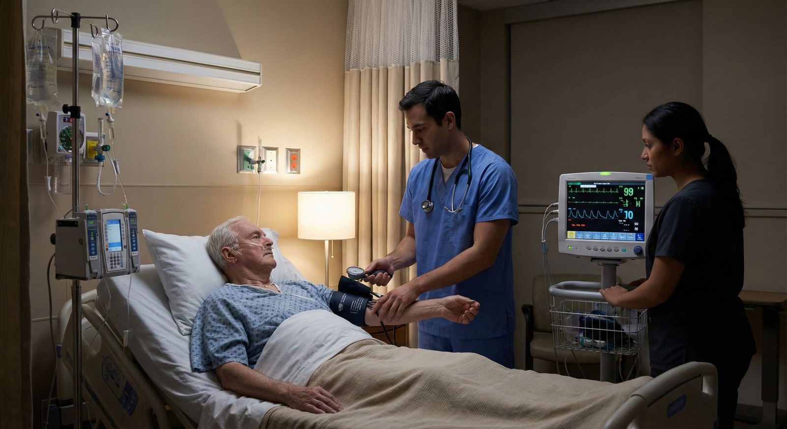
The human body conserves water for dozens of reasons that have nothing to do with systemic dehydration. Postoperative surgical patients experience a surge of aldosterone and antidiuretic hormone (ADH) as a normal physiologic reaction to surgical tissue trauma. Their kidneys are holding onto sodium and water by design. If the patient is normotensive, comfortably resting, and clinically euvolemic, that transient dip in urine output overnight is often normal physiology working as intended.
When you flood these patients with saline to make the chart look nice, you aren't fixing a hydration deficit. You are diluting their hematocrit, stretching their right ventricle, and swelling their interstitial tissues.
Urine output falls due to a wide variety of underlying causes:
- Mechanical obstruction or retention (a kinked Foley, a clogged lumen, or post-void retention)
- Measurement and charting errors (an unrecorded drainage dump or a patient walking to the bathroom without telling nursing)
- Impaired perfusion (cardiogenic shock, distributive shock, or elevated intra-abdominal pressure)
- Intrinsic kidney injury (acute tubular necrosis, interstitial nephritis, or contrast-induced injury)
- Medication effects (diuretics wearing off, ACE inhibitors altering glomerular hemodynamics, or anticholinergics causing urinary retention)
The clinical evidence is clear: blind fluid administration in non-responsive oliguric patients does not restore kidney function. It simply worsens fluid creep.
What to Check First: A Rapid Oliguria Triage Framework
When that 3 AM page hits, run through a structured evaluation before reaching for crystalloid fluids.
1. Verify the Measurement
First, ensure the problem isn't mechanical or human error. Is the catheter actually patulous? I've lost count of how many times "oliguria" was cured by stripping a kinked catheter tube or flushing a clogged port that was occluded by blood or sediment. If the patient doesn't have a catheter, order a bedside bladder scan immediately. A patient sitting with 800 mL of urine trapped behind a spasms-plagued prostate doesn't need a liter of normal saline, they need a Foley catheter.
2. Assess Perfusion and Hemodynamics
Walk to the bedside. Check the blood pressure, heart rate, and mean arterial pressure (MAP). Touch the patient's extremities. Are they warm and well-perfused, or cold, mottled, and clammy? Examine the jugular venous pressure (JVP). Listen to the lung fields. Check the intake and output log for the past 48 hours, is the patient net positive 6 liters since surgery? If so, true hypovolemia is down at the bottom of your differential.
3. Review the Clinical Context
Look at the med list and surgical notes. Did they receive IV contrast in the catheterization lab yesterday? Did someone start an NSAID, an ACE inhibitor, or double the dose of vancomycin? Did the intraoperative team leave them with an open abdomen or high peak airway pressures suggestive of elevated intra-abdominal pressure? Frame the drop in output against their baseline organ function.
When Fluids Help, and When They Hurt
Fluids are a drug with a narrow therapeutic window. They increase urine output under one specific physiological condition: when low renal blood flow is driven by true intravascular volume depletion or early unresuscitated distributive shock, and the heart is sitting on the steep, fluid-responsive portion of the Frank-Starling curve.
Once a patient shifts to the flat top of that cardiac output curve, extra volume offers no hemodynamic benefit. Instead, cardiac filling pressures skyrocket. Venous pressure backs up straight into the renal veins.
Because the kidneys are encapsulated organs, high renal venous pressure increases interstitial pressure within the kidney, reducing the net pressure gradient across the glomerulum. Giving excess fluids to a hypervolemic patient can actually decrease their glomerular filtration rate (GFR) and worsen oliguria.
Red Flags Against Giving Boluses:
- Peripheral edema, sacral edema, or anasarca
- Crackles on lung exam or new or worsening oxygen requirements
- Known baseline heart failure with reduced ejection fraction or end-stage liver disease with ascites
- Cumulative positive fluid balance exceeding 2-3 liters over their admission
- Complete anuria (which suggests total obstruction, vascular catastrophic blockage, or severe cortical necrosis)
If you decide a fluid challenge is appropriate, frame it as a strict diagnostic trial: give a small fluid challenge (such as 250-500 mL of balanced crystalloid over 15 to 30 minutes) and reassess. Did the heart rate drop? Did MAP improve? Did urine output pick up within the next hour? If the answer is no, stop giving fluids. Repeating the same ineffective intervention while expecting a different renal outcome is a classic training trap.
The On-Call Workup That Actually Changes Management
You don't need to order every lab on the panel at 3 AM, but targeted diagnostics save lives.

High-Yield Bedside Tools
- Bedside Bladder Scanner: The single most useful diagnostic tool for nocturnal oliguria. It differentiates post-renal obstruction from intra-renal or pre-renal causes in five seconds.
- Point-of-Care Ultrasound (POCUS): If you are trained, check the inferior vena cava (IVC) collapsibility, look for lung B-lines (suggesting pulmonary edema), or evaluate for hypervolemic venous congestion using VEXUS concepts.
- Basic Metabolic Panel: Check the blood urea nitrogen (BUN), serum creatinine, and potassium levels. Look at the creatinine trend rather than a single isolated value. A jump in creatinine confirms true acute kidney injury.
- Urinalysis & Urine Electrolytes: A clean urinalysis suggests pre-renal physiology or hemodynamic changes. Granular casts suggest acute tubular necrosis. Note: Fractional excretion of sodium (FeNa) is unreliable if the patient received loop diuretics in the preceding 12-24 hours; use fractional excretion of urea (FeUrea) instead.
When to Call Your Fellow or Attending
Do not suffer in silence when the clinical picture deteriorates. Escalate early if you encounter:
- Sudden complete anuria (0 mL over 2 hours despite an open, non-obstructed catheter)
- Severe refractory hyperkalemia or severe metabolic acidosis tied to oliguric acute kidney injury
- New or worsening respiratory failure or severe pulmonary edema following fluid administration
- Oliguria in the context of persistent, refractory septic or cardiogenic shock requiring new or escalating vasoactive drips
Putting It Together: A Practical Script for Residents on Call
When you get that overnight call for low urine output, use this simple mental algorithm:
- Verify: "Is the catheter clear, or does a bladder scan show urinary retention?"
- Assess: "Is the patient hypotensive or hypoperfused? Or are they normotensive, comfortably resting, and fluid-overloaded?"
- Differentiate: "Is this acute kidney injury, mechanical obstruction, normal post-op neuroendocrine response, or over-diuresis?"
- Act: "Does the underlying physiology truly warrant a fluid bolus, or should I hold fluids, adjust medications, or simply monitor the patient?"

Clinical Scenarios in Practice
- Scenario A: A Post-Op Day 1 patient following an abdominal procedure has a urine output of 18 mL/hr overnight. MAP is 82 mmHg, heart rate is 74, lungs are clear, and they are net positive 4 liters from the operating room. Action: Perform a bladder scan to confirm catheter patency. If clear, hold fluids, reassure nursing, and reassess in two hours. Do not give a fluid bolus to "fix" normal post-surgical endocrine stress response.
- Scenario B: An ICU patient with severe intra-abdominal sepsis has a MAP drop to 52 mmHg, HR increases to 125, skin is mottled, and urine output drops to 5 mL/hr. Action: Evaluate overall fluid responsiveness. Initiate or titrate vasopressors to restore adequate renal perfusion pressure (target MAP ≥ 65 mmHg) rather than relying exclusively on continuous crystalloid loading.
- Scenario C: A patient admitted with acute decompensated heart failure is receiving intravenous furosemide. Urine output drops off after initial diuresis, and creatinine rises from 1.1 to 1.5 mg/dL. Action: Assess perfusion status. If perfusing well, the elevated creatinine and low urine output may reflect temporary hemodynamically mediated drops in GFR, not a reason to give back the fluid you just diuresed.
Shift your mindset away from viewing urine output as a numerical goal to hit with an IV bag. Treat the underlying physiology, protect the patient from fluid overload, and use data, not reflexes, to guide your care on call.
Key Takeaways
- Oliguria is a symptom, not a diagnosis. Dehydration is only one of many underlying causes for low urine output.
- Verify before treating. Always rule out mechanical obstruction, catheter kinks, charting errors, and post-void retention using a bladder scan before ordering interventions.
- Fluid boluses can worsen renal function. In fluid-overloaded or non-responsive patients, excessive IV crystalloid increases renal venous pressure and decreases GFR.
- Reassess after every intervention. If you give a targeted fluid challenge, look for explicit clinical improvements in MAP, perfusion, and output. If it fails, stop giving fluids.
- Escalate severe cases early. In cases of absolute anuria, refractory hyperkalemia, or worsening pulmonary edema, contact senior physicians early.